

Translate this page into:
Zinner syndrome: A rarer variant of a rare syndrome
*Corresponding author: Amit Gupta, Department of Radiodiagnosis and Interventional Radiology, All India Institute of Medical Sciences, New Delhi, Delhi, India. amit.aiims2014@gmail.com
-
Received: ,
Accepted: ,
How to cite this article: Ravisandhiran B, Das CJ, Gupta A. Zinner Syndrome: A rarer variant of a rare syndrome. Case Rep Clin Radiol. doi: 10.25259/CRCR_147_2023
Abstract
Zinner syndrome, characterized by the combination of absent unilateral kidney, obstructed ejaculatory duct on the same side, and cysts in the seminal vesicle, represents a rare congenital malformation of the mesonephric duct. Its symptoms are often vague, ranging from urinary discomfort to painful ejaculation and infertility, making it difficult to diagnose without imaging. In this report, we discuss a less common form of Zinner syndrome identified through diagnostic imaging.
Keywords
Zinner syndrome
Seminal vesicle cysts
Renal agenesis

INTRODUCTION
Zinner syndrome, a seldom-seen congenital anomaly of the mesonephric duct, is delineated by a distinct triad: Cysts in the seminal vesicle, blockage of the ejaculatory duct, and the absence of the kidney on the same side, with an occurrence estimated at 200 cases/1 million population.[1,2] Herein, we describe a less common variant of Zinner syndrome identified through diagnostic imaging.
CASE REPORT
A 21-year-old male presented to the urology outpatient department with complaints of dysuria and increased frequency. Physical examination and laboratory work-up were unremarkable. The patient was referred to radiology for computed tomography (CT) urography for further causal evaluation. CT revealed absence of the left kidney and presence of a hypodense cystic attenuation structure in the right pelvis posterior to the urinary bladder [Figure 1]. Corroborative ultrasound (USG) and magnetic resonance imaging (MRI) showed presence of the right seminal vesicle cysts and non-visualized likely hypoplastic left seminal vesicle [Figure 2]. Possibility of a variant of Zinner syndrome was kept. Subsequent semen analysis showed azoospermia and the patient was planned for transurethral resection of the ejaculatory duct.

- (a) Venous phase CT axial section shows agenesis of left kidney with empty left renal fossa containing displaced bowel loops (asterisk). (b) Axial pelvic section shows elongated cystic attenuation structure (white arrow) posterior to the urinary bladder (UB) in right paramedian location.
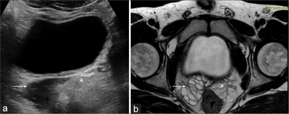
- (a) Pelvic USG shows well defined elongated anechoic cystic structure (white arrow) in retrovesical and right periprostatic location. (b) Axial T2-weighted MRI image shows enlarged lobulated right seminal vesicle entirely replaced by multiple hyperintense cysts (white arrow) with non-visualisation of left seminal vesicle (asterisk in a and b).
DISCUSSION
Zinner syndrome is a rare congenital defect occurring due to failure of interaction between ureteric bud and metanephric blastema affecting the development of their derivatives.[1,3] This condition typically manifests in the second and third decades of life with symptoms ranging from such as dysuria and hematuria to recurrent infections, painful ejaculation, and infertility.[1,2]
The hallmark of Zinner syndrome consists of cysts in the seminal vesicle, blockage of the ejaculatory duct, and ipsilateral renal agenesis. The presence of a seminal vesicle cyst on the opposite side of a missing kidney represents an exceedingly uncommon variant of Zinner syndrome, with only four such cases reported so far.[4] Even rare variants involve agenesis or hypoplasia of contralateral seminal vesicle or either testis[5] [Figure 3].

- (a) Schematic diagram showing anatomy in the characteristic triad of Zinner syndrome. (b) Variant of Zinner syndrome reported in the index case. (c) Another reported rarer variant of Zinner syndrome. Figure illustrated by Bharathi Ravisandhiran (co-author).
Diagnosis of Zinner syndrome is primarily reliant on imaging. Ultrasound is the preferred initial screening modality and can readily identify the unilateral renal agenesis along with anechoic spaces in the pelvis secondary to seminal vesicle cysts and obstructed ejaculatory ducts. MRI stands as the superior imaging technique for examining pelvic structures and identifying genitourinary anomalies, offering a clear view of the cysts’ origins, and assisting in pre-surgical planning.[2,5] The utility of CT in visualization of pelvic cysts is limited. However, as in the index case, all cases of renal agenesis in male patients should be carefully scrutinized for the presence of hypodense seminal vesicle cysts in the lower pelvic CT sections which can be further confirmed on USG or MRI in case of a suspicion.
Treatment for asymptomatic cases tends to be conservative, while surgical interventions such as ejaculatory duct resection or vesiculectomy are options for those experiencing symptoms, often resulting in significant improvement and potentially restoring fertility.[1,3,4]
CONCLUSION
Zinner syndrome represents a unique congenital urogenital anomaly, often presenting with vague symptoms. Nevertheless, definitive diagnosis is achievable through characteristic imaging findings of Zinner syndrome and its rarer forms, guiding the effective treatment of affected individuals.
TEACHING POINTS
Zinner Syndrome, characterized by ejaculatory duct obstruction, seminal vesicle cysts, and ipsilateral renal agenesis, results from a defect during embryogenesis of mesonephric duct in a male child.
It is usually asymptomatic, however, can have myriad presentations with symptoms ranging from dysuria to painful ejaculation and infertility requiring prompt management.
Diagnosis relies heavily on imaging, with ultrasound as the initial step and MRI as the definitive method for evaluating the condition.
MCQs
-
Which is the best imaging modality for diagnosis of Zinner syndrome?
USG
CT
MRI
Radiographs
Answer Key: c
-
Zinner syndrome is characterized by presence of which of these anatomical abnormalities?
Aortic aneurysm
Hepatic cysts
Urethral stricture
Seminal vesicle cysts
Answer Key: d
-
On a radiological image, a seminal vesicle cyst in Zinner syndrome is most commonly located adjacent to which structure?
Bladder
Rectum
Prostate gland
Testis
Answer Key: c
Ethical approval
Institutional Review Board approval is not required.
Declaration of patient consent
The authors certify that they have obtained all appropriate patient consent.
Conflicts of interest
There are no conflicts of interest.
Use of artificial intelligence (AI)-assisted technology for manuscript preparation
The authors confirm that there was no use of artificial intelligence (AI)-assisted technology for assisting in the writing or editing of the manuscript and no images were manipulated using AI.
Financial support and sponsorship
Nil.
References
- Zinner’s syndrome, radiological diagnosis for a rare syndrome with non-specific clinical presentation: Case report. Egypt J Radiol Nucl Med. 2020;51:217.
- [CrossRef] [Google Scholar]
- Zinner syndrome: A unique triad of mesonephric duct abnormalities as an unusual cause of urinary symptoms in late adolescence. Indian J Urol. 2010;26:444-7.
- [CrossRef] [PubMed] [Google Scholar]
- Unique association of multiple seminal vesicle cysts with contralateral renal agenesis: A rare variant of Zinner syndrome. Cureus. 2017;9:e1415.
- [CrossRef] [Google Scholar]
- A rare case of Zinner syndrome with unilateral testicular agenesis and aberrant course of left common iliac artery. Egypt J Radiol Nucl Med. 2023;54:99.
- [CrossRef] [Google Scholar]


